
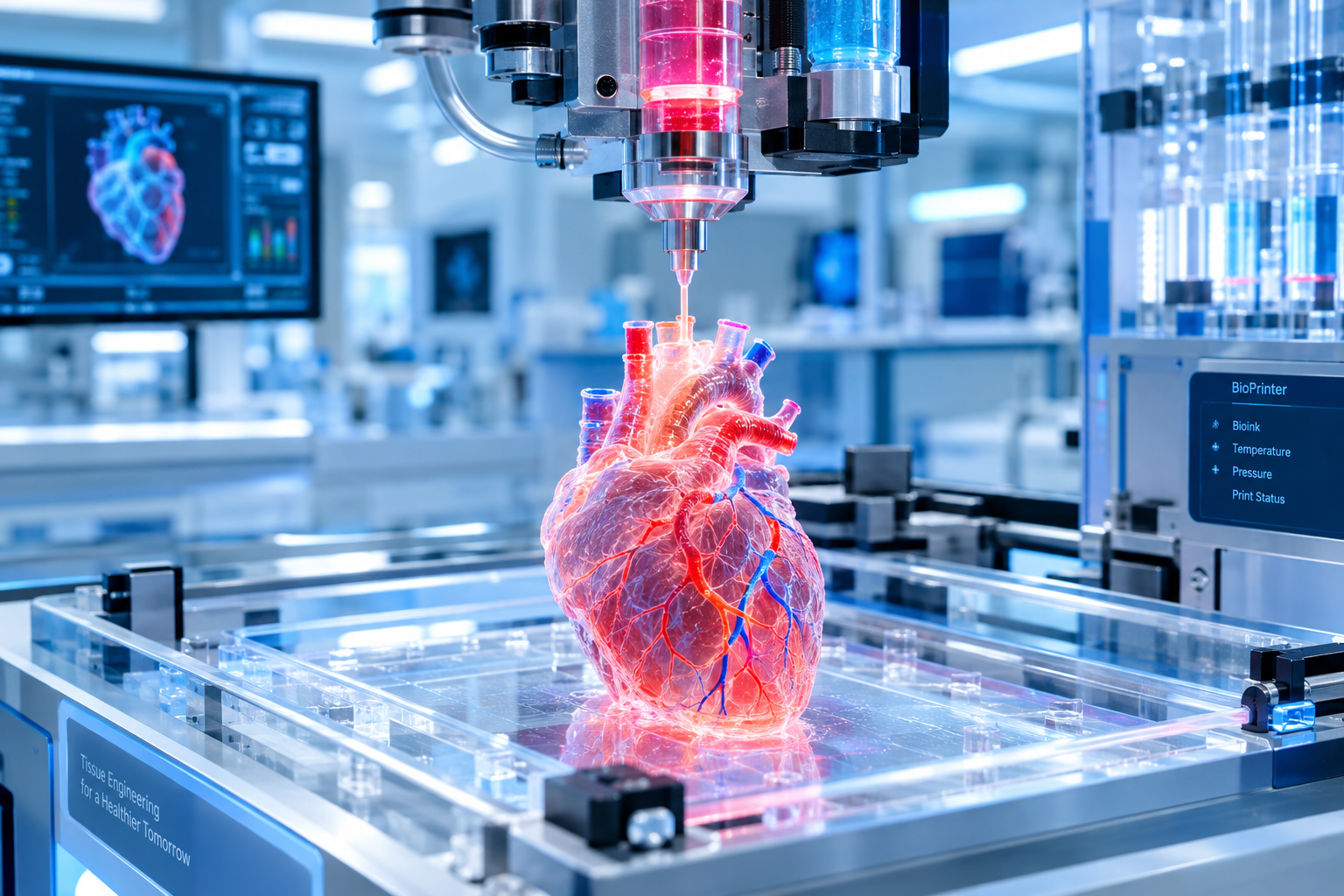
Every single day, across hospitals worldwide, a silent and heartbreaking race against time plays out. Hundreds of thousands of patients suffering from end-stage organ failure sit on national organ transplant waiting lists. In the United States alone, an average of 17 people die every day simply waiting for an available donor organ, while a new patient is added to the national registry every eight minutes.
For decades, medicine's only answer has been donor transplantation-a system constrained by extreme organ shortages and fraught with the constant risk of biological rejection. But at the intersection of cell biology, materials science, and advanced robotics, a technological revolution is underway: 3D Bioprinting.
What Is 3D Bioprinting?
Traditional 3D printing (additive manufacturing) builds inanimate geometric objects by extruding layers of molten plastic, resin, or metal powder. 3D Bioprinting adapts this layer-by-layer methodology to living biology. Instead of industrial thermoplastics, bioprinters utilize bioinks-formulations composed of living human cells suspended inside cytocompatible hydrogels that mimic the natural extracellular matrix (ECM) of human tissue.
The 4-Stage Workflow of Printing a Living Organ
Constructing a complex, vascularized human organ involves a sophisticated four-stage bio-engineering pipeline:
1. Patient-Specific Cellular Harvesting (iPSCs)
To eliminate any risk of immune rejection, the process begins with the patient's own cells. A small skin biopsy or blood draw is harvested, and the cells are genetically reprogrammed into induced Pluripotent Stem Cells (iPSCs). These master cells possess the extraordinary ability to differentiate into any cell type in the human body-whether cardiac myocytes (heart muscle), hepatocytes (liver cells), or renal podocytes (kidney filters).
2. Bioink Formulation & Scaffold Synthesis
The differentiated cells are multiplied by the billions inside bioreactors and combined with specialized biocompatible hydrogels (derived from alginate, collagen, hyaluronic acid, and gelatin-methacryloyl). The bioink must possess precise rheological properties: it must flow smoothly through microscopic printer nozzles under shear stress without damaging delicate cell membranes, and instantly cross-link into a rigid, self-supporting structure upon deposition.
3. High-Resolution Multi-Material Printing
Using advanced multi-nozzle pneumatic extrusion or stereolithographic laser projectors, the bioprinter executes a digital CAD model reconstructed from the patient's high-resolution MRI or CT scan. The printer deposits distinct cell types, structural scaffolding, and microscopic channel pathways simultaneously with micron-level precision.
4. Bioreactor Maturation & Functional Vascularization
A freshly printed organ is not immediately ready for implantation; it is essentially a fragile cellular construct. The organ is transferred into a biomimetic bioreactor-a sealed chamber that supplies warm, oxygenated nutrient broth and applies physical stimuli (such as rhythmic electrical pulses for cardiac tissue or mechanical perfusion for blood vessels) until the cells self-organize, form intercellular gap junctions, and begin beating or filtering fluids as a unified organ.
Overcoming the Ultimate Hurdle: The Vascularization Challenge
For years, the Achilles' heel of tissue engineering was vascularization. Human cells cannot survive if they are located more than 200 microns (the thickness of two human hairs) away from an oxygen-delivering capillary. Without a blood supply, the interior cells of thick 3D-printed tissues rapidly suffocate and die of necrosis.
Researchers have conquered this limitation through sacrificial templating and two-photon laser polymerization. Bioprinters print intricate vascular networks using temporary, water-soluble fugitive inks (like Pluronic F-127). Once the surrounding tissue matrix solidifies, the fugitive ink is washed away, leaving hollow, branching capillary channels that are seeded with human endothelial cells to create functional, leak-free blood vessels.
Clinical Milestones on the Near Horizon
- Living Skin Grafts for Burn Victims: Mobile bioprinters can already scan burn wounds in surgical theaters and print customized skin layers directly onto patients in real time.
- Vascularized Meniscus & Cartilage Implants: Printed knee cartilage constructs have successfully completed clinical trials, restoring joint mobility without requiring metallic prosthetic replacements.
- Micro-Liver Lobules for Pharmaceutical Testing: 3D-printed mini-livers are already replacing animal testing in toxicology trials, speeding up drug discovery pipelines worldwide.
- Full Solid Organ Replacement: Functional, transplanted bioprinted kidneys and hearts are advancing rapidly through pre-clinical large-animal models, with initial human compassionate-use trials anticipated before the end of the decade.
Ethical and Regulatory Dimensions of Bioprinting
As 3D bioprinting transitions into mainstream medicine, regulatory frameworks (such as FDA Good Tissue Practice guidelines) are evolving to standardize quality control for living therapies. The ability to print organs on demand also eliminates the illicit black market for human organs, establishing an ethical, equitable framework for global healthcare.
A World Free From the Organ Shortage Crisis
3D Bioprinting promises to transform organ failure from an agonizing death sentence into a manageable, elective medical procedure. When an organ fails, doctors will simply pull up a patient's medical scan, load their personalized stem-cell bioink into a printer, and synthesize a pristine, genetically identical replacement organ on demand. The end of the transplant waiting list is no longer a utopian dream; it is the near-future reality of regenerative medicine.
The Roadmap from Lab Bench to Operating Room
| Tissue / Organ Type | Complexity Level | Current Clinical Stage | Projected Routine Implantation |
|---|---|---|---|
| Skin & Cartilage | Low (Avascular / Thin) | Active Human Clinical Trials | 2026 - 2028 |
| Blood Vessels & Trachea | Moderate (Hollow tubular) | Pre-clinical / Compassionate use | 2028 - 2030 |
| Kidneys & Liver Lobules | High (Complex filtration) | Large animal pre-clinical trials | 2031 - 2035 |
| Four-Chambered Heart | Extreme (Synchronized electromechanics) | Early functional bio-prototyping | 2035+ |
Frequently Asked Questions (FAQ)
1. How long does it take to 3D-print a human organ?
Printing the physical cellular scaffold takes from 4 to 24 hours depending on organ size. However, maturing the printed construct inside a biomimetic bioreactor to achieve full biological function takes 2 to 6 weeks.
2. Will bioprinted organs be affordable?
While initial clinical prototypes will carry high costs, the elimination of lifelong immunosuppressant medication (20,000-40,000/year) and automated bioreactor scaling will make bioprinting significantly cheaper than lifelong dialysis or organ failure management.
Case Study: The 3D-Bioprinted Micro-Kidney Revolution
Chronic Kidney Disease (CKD) accounts for massive global healthcare expenditure, with millions of patients reliant on hemodialysis. Researchers have successfully bioprinted human kidney organoids complete with functional nephrons, glomeruli, and proximal tubules. When connected to fluidic flow circuits, these micro-printed tissues successfully filter metabolic waste, reabsorb electrolytes, and respond to nephrotoxic medications identically to native human kidneys, bringing fully functional whole-organ kidney printing closer to clinical surgery.
Biocompatible Cross-Linking and Photo-Polymerization
To ensure that bioprinted tissues maintain structural integrity inside the human body without collapsing under physical blood pressure, bioengineers utilize ultraviolet and blue-light photo-cross-linkable hydrogels (such as GelMA). By shining targeted laser patterns during deposition, the hydrogel polymers cross-link within milliseconds, forming an elastic yet durable biological extracellular matrix that native host blood vessels can integrate with.